[最も欲しかった] hepg2 oil red o staining 119252-Oil red o staining hepg2 cells protocol
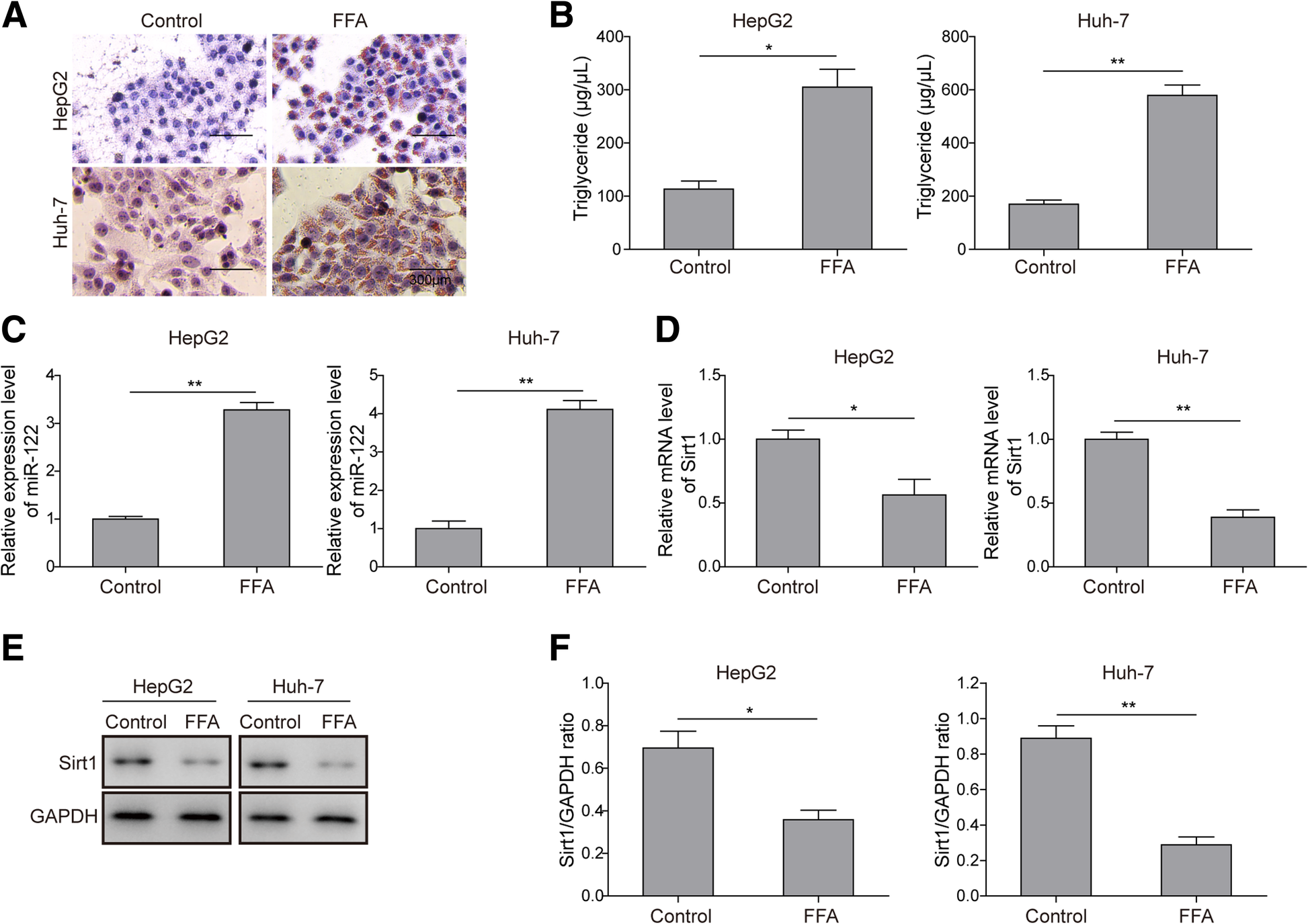
Mir 122 Promotes Hepatic Lipogenesis Via Inhibiting The Lkb1 Ampk Pathway By Targeting Sirt1 In Non Alcoholic Fatty Liver Disease Molecular Medicine Full Text
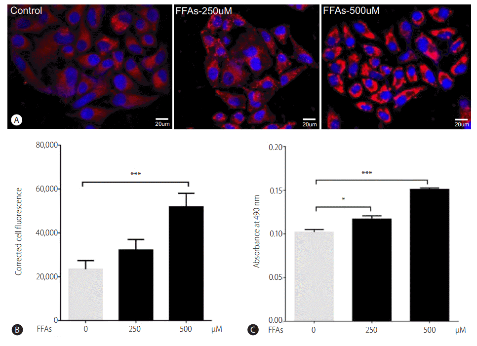
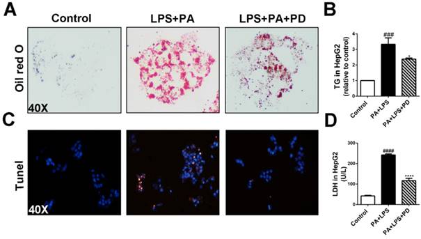
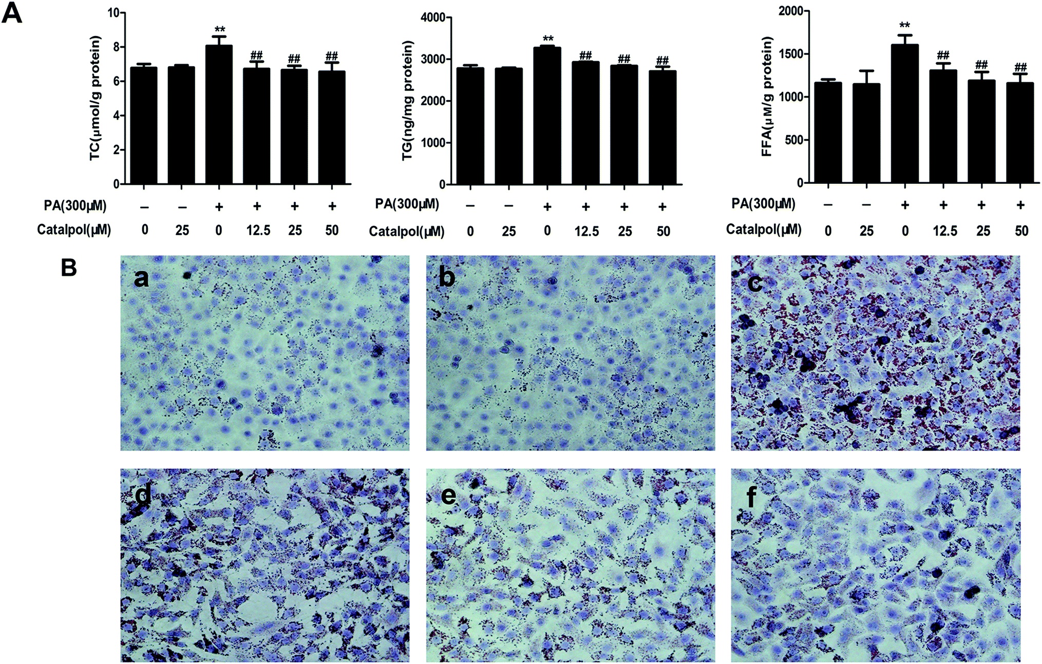
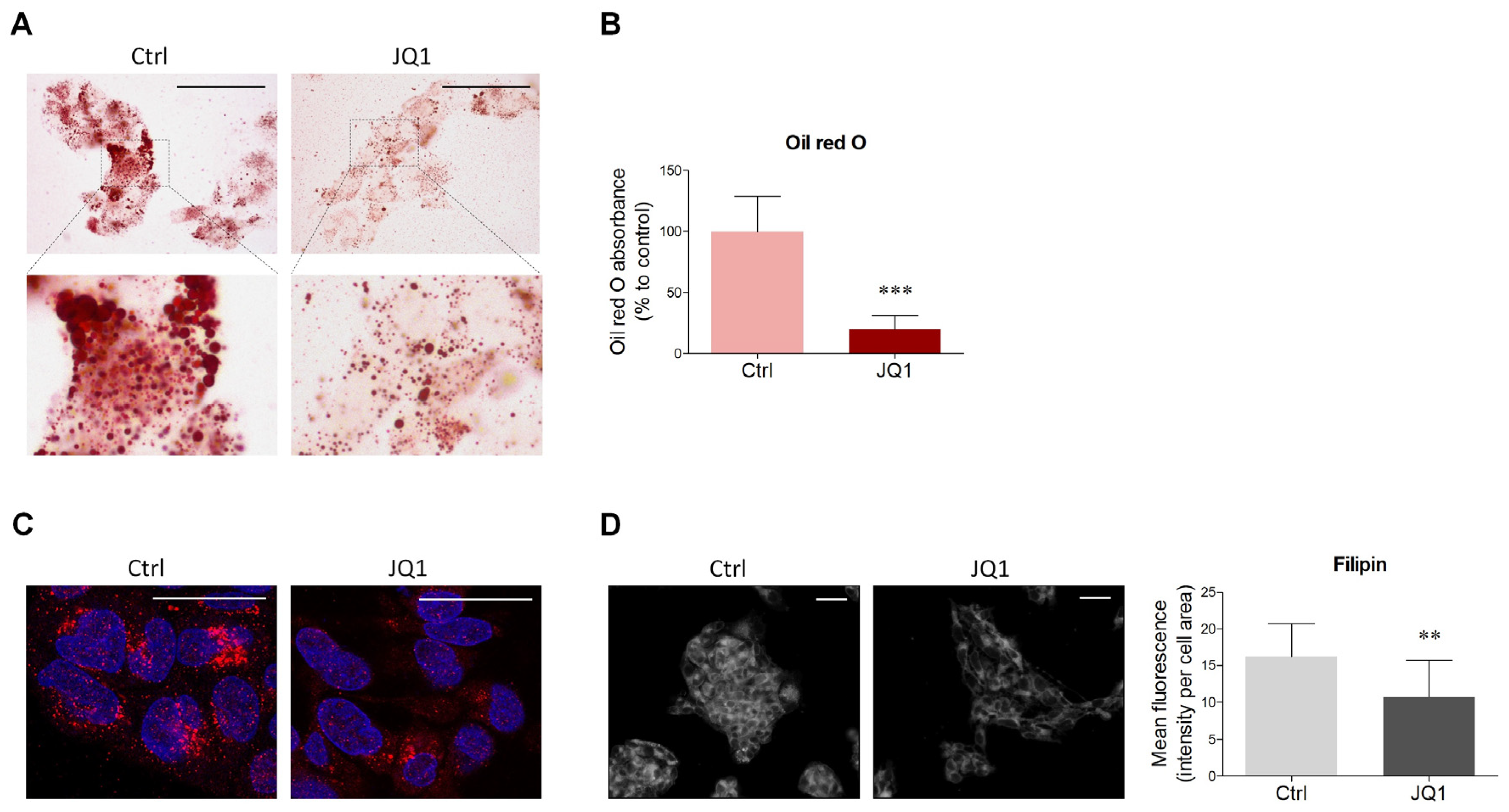
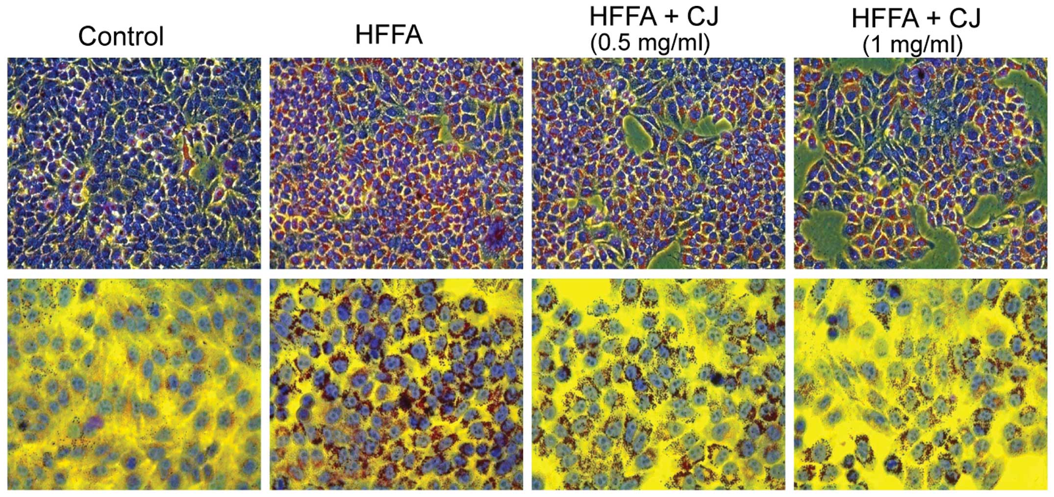
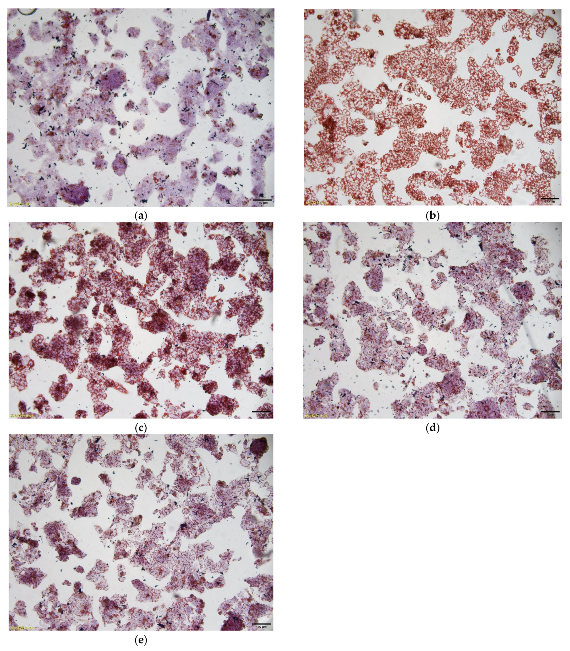
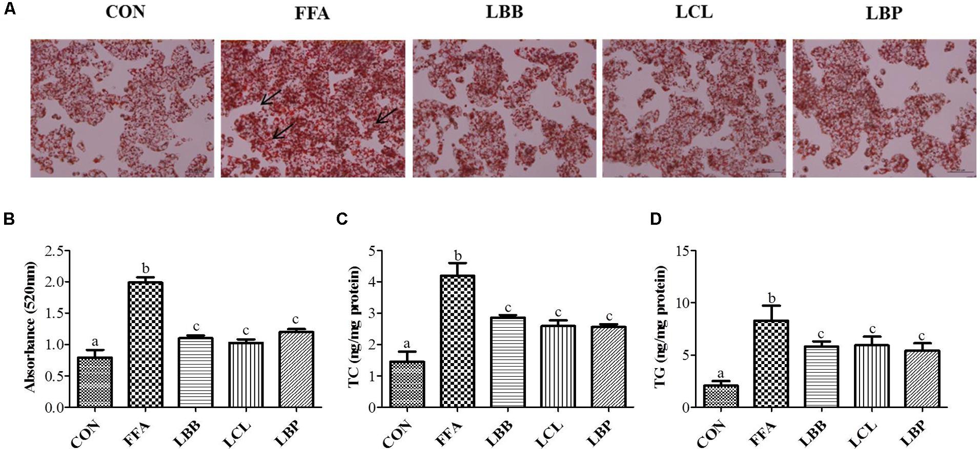
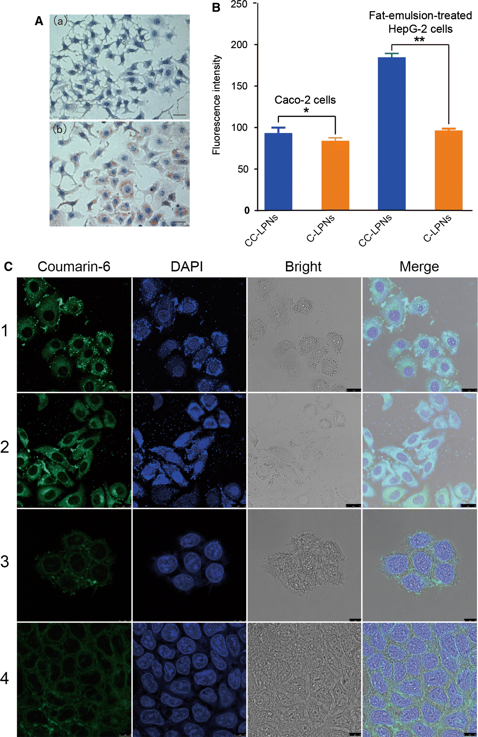
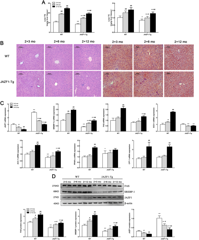
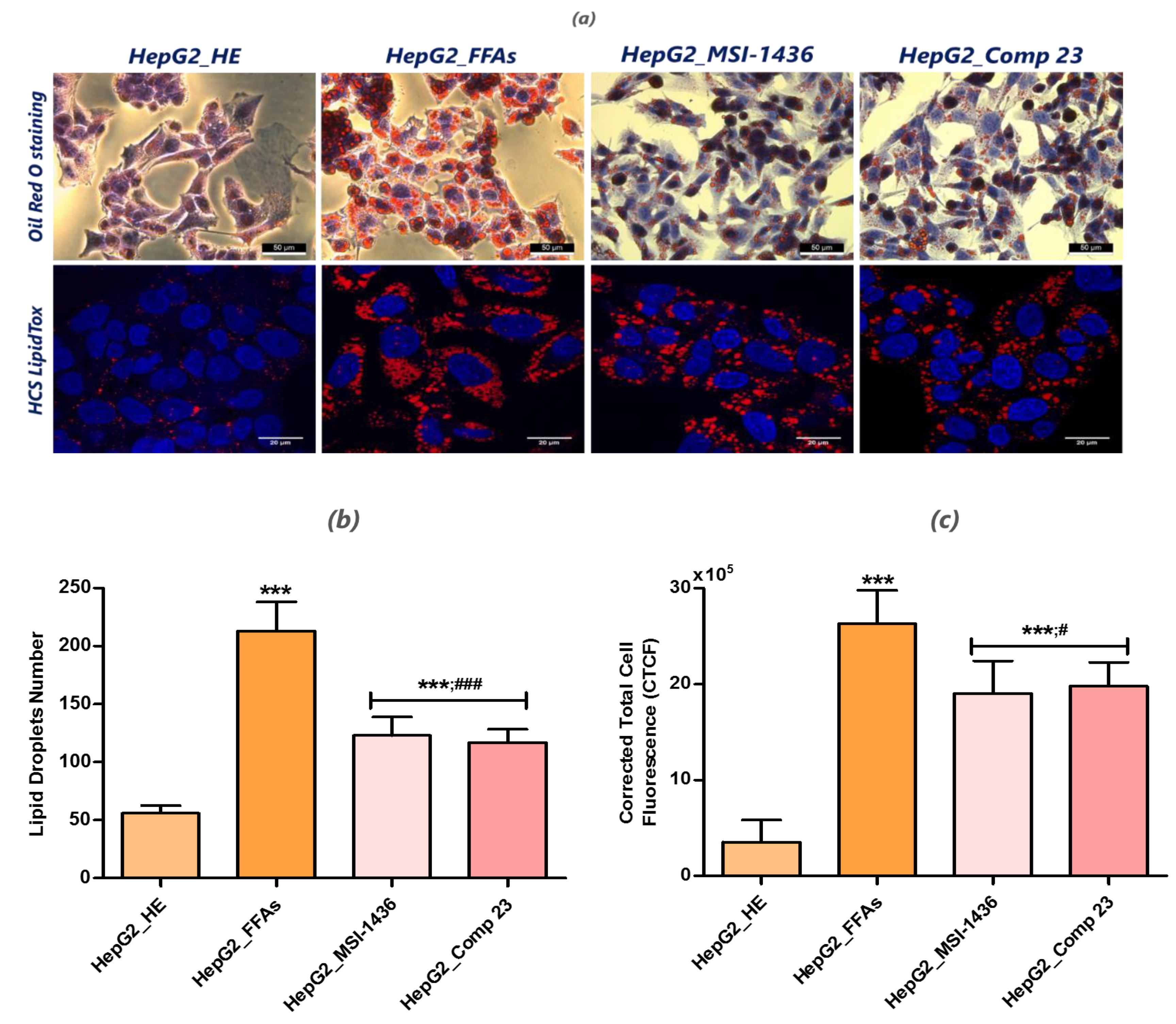
The Oilred O staining result showed obvious difference of the existence of lipid droplets between the FFAexposed HepG2 cells and control, and the concentration of TG and TC in FFAexposed HepG2 cells was 86 ng/10 4 cells and 06 nM/104 cells, which were significantly different with control croup (p < 05)Preparing oil red o stain • Prepare the stock solution by weighing out 300 mg of oil red o powder and adding this to 100 ml of 99% isopropanol This solution is stable for one year from the date on which it is prepared • In the fume hood, mix 3 parts (30 ml) of oil red o stock solution with 2 parts ( ml) deionized water and
Oil red o staining hepg2 cells protocol
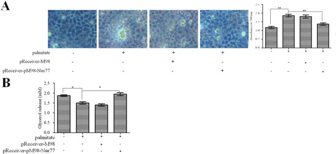
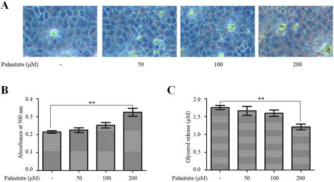
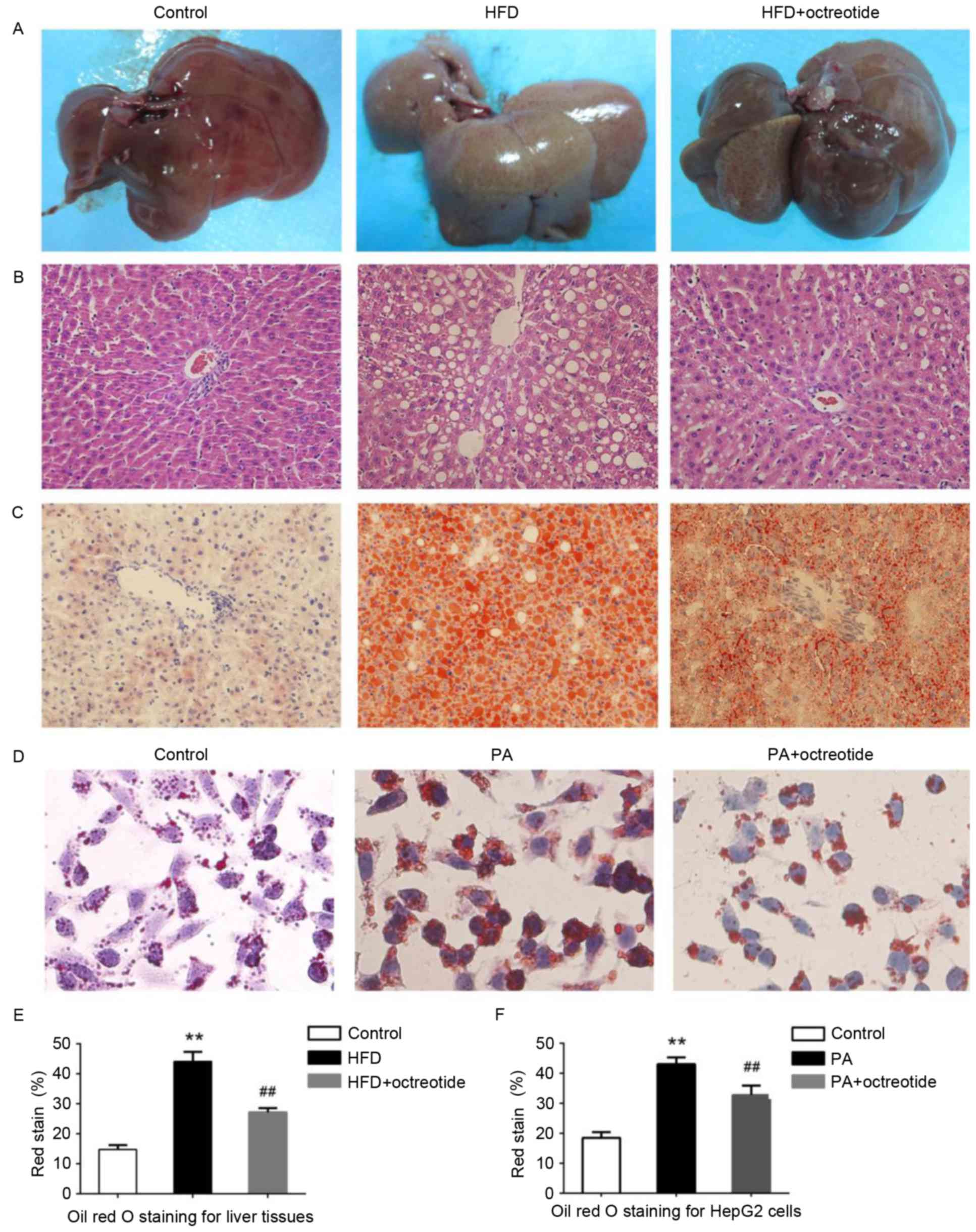
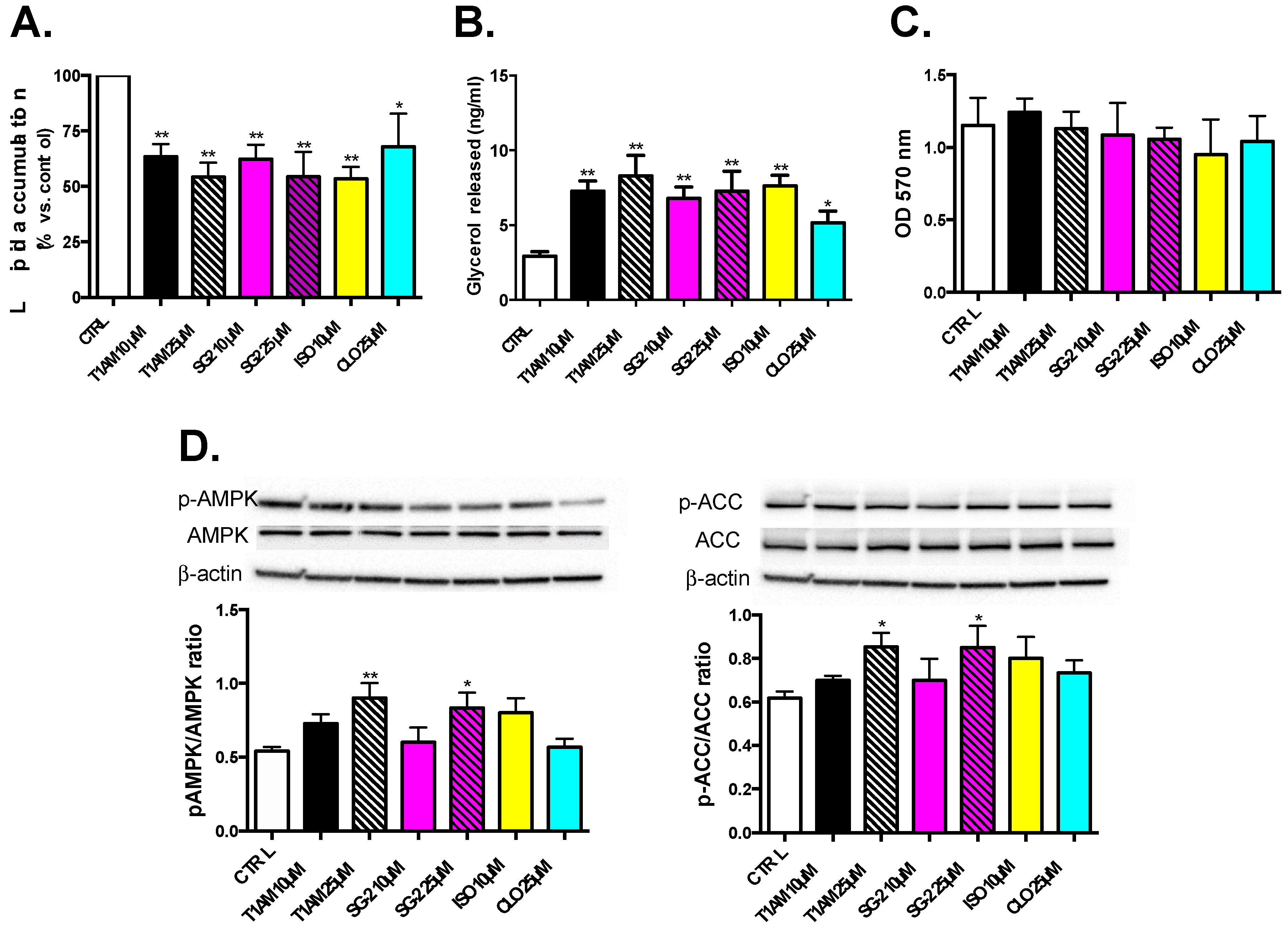
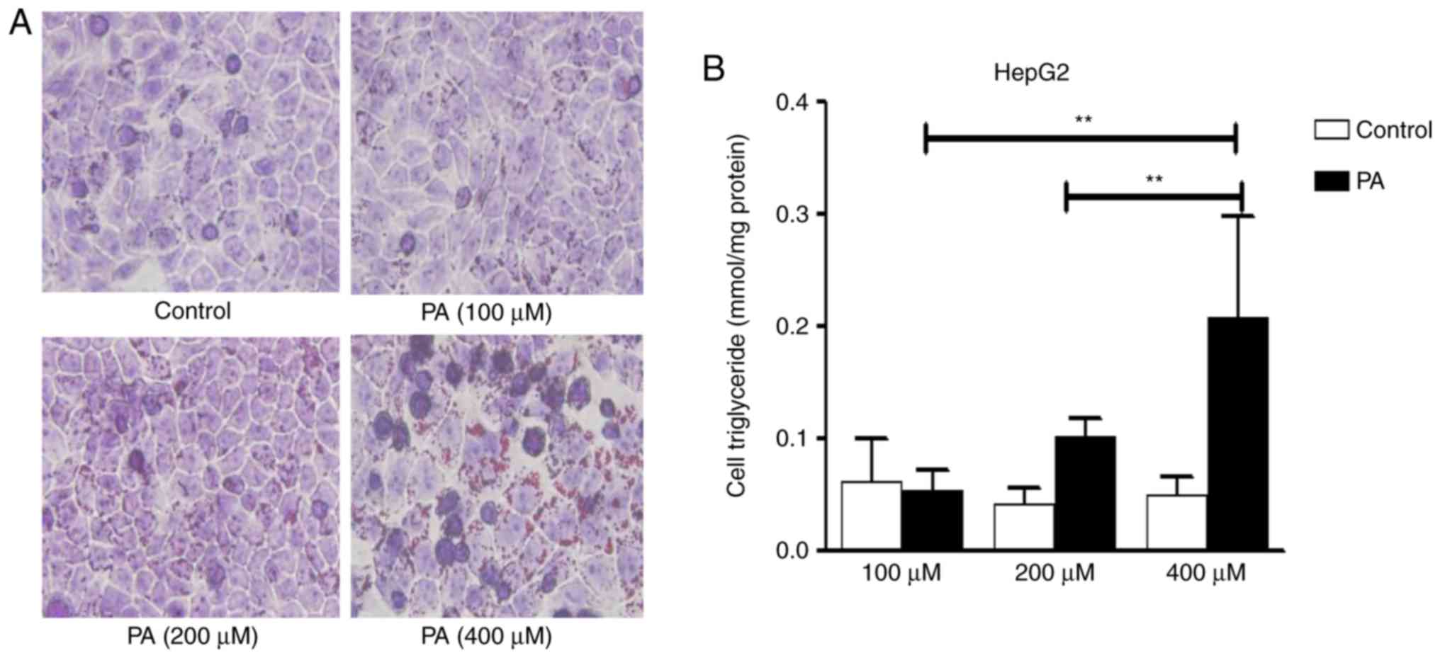
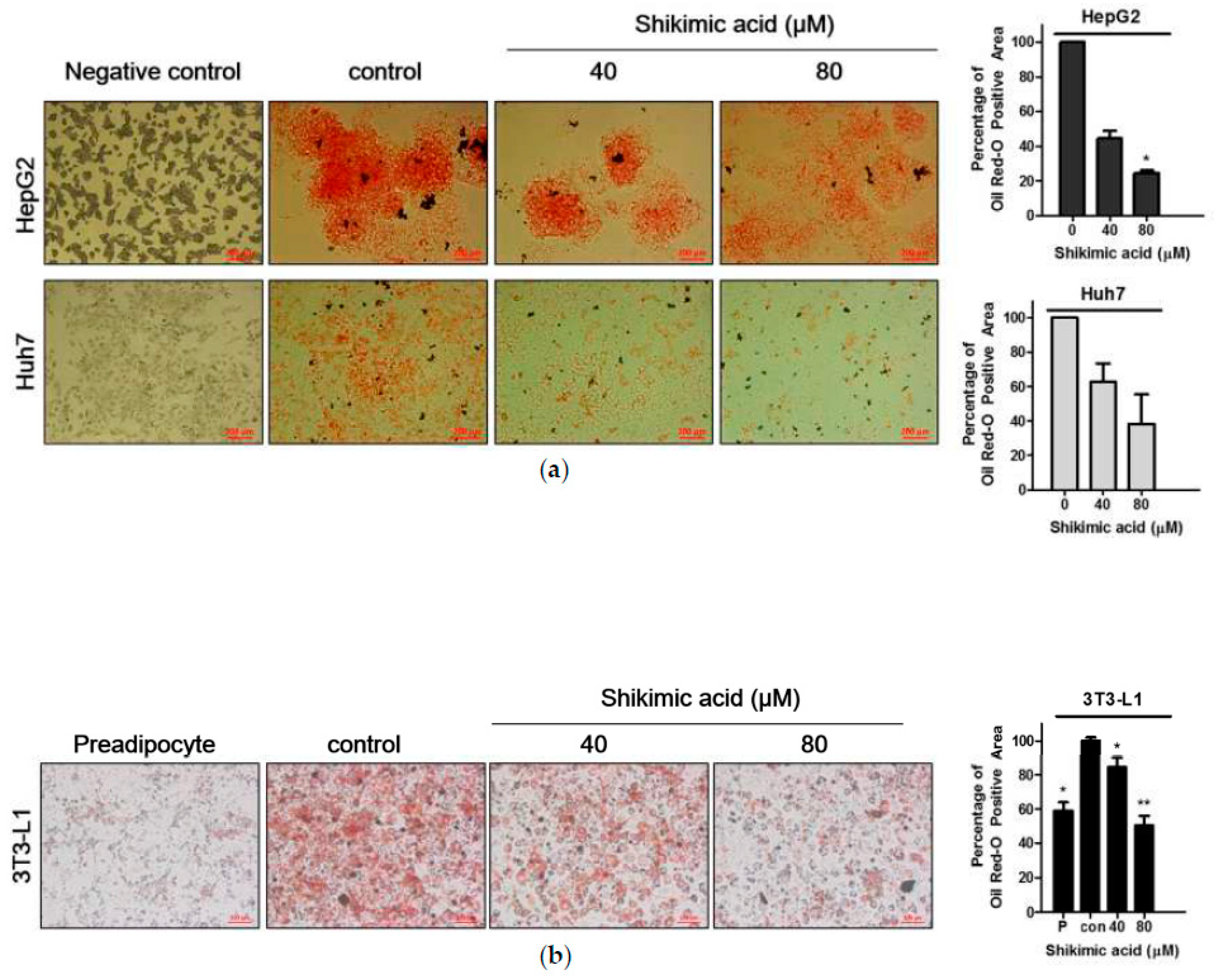
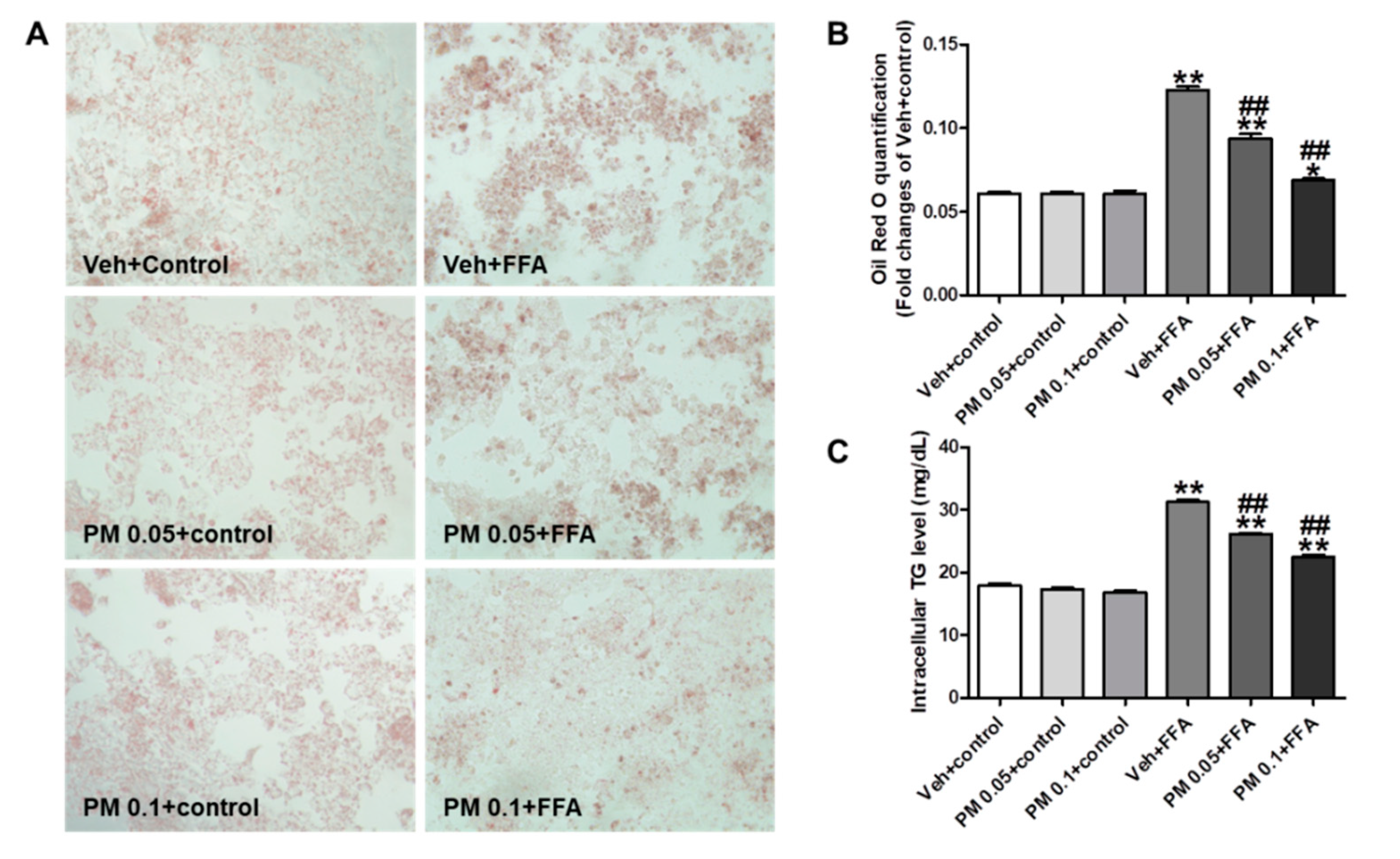
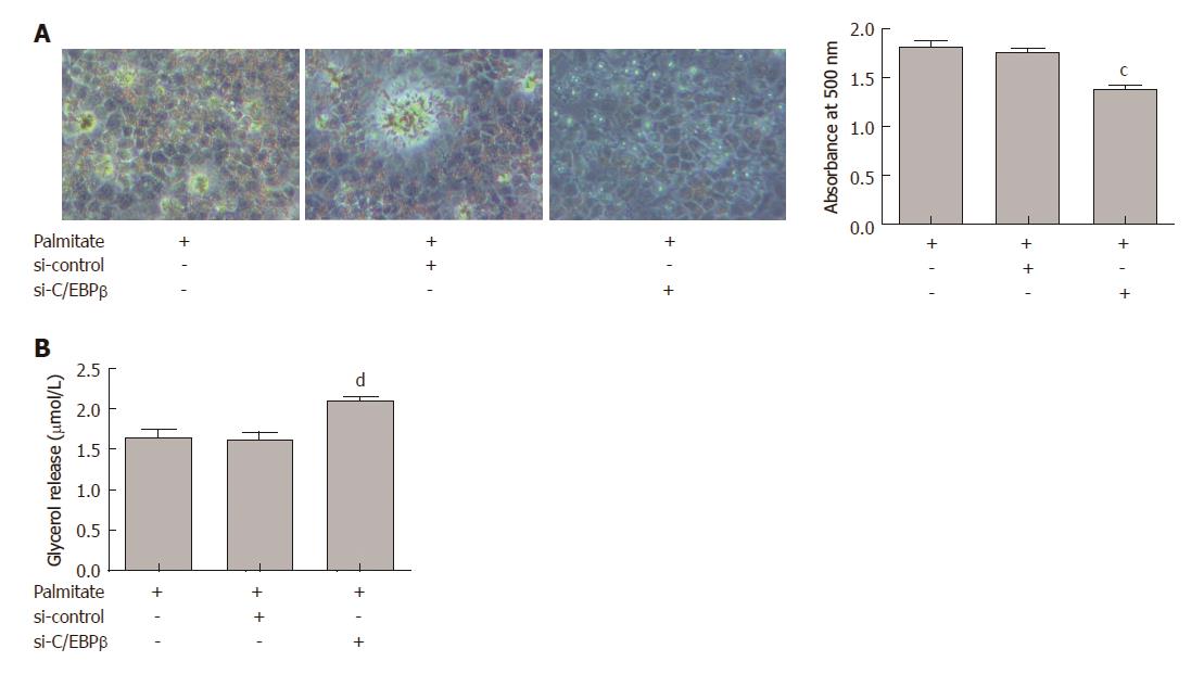
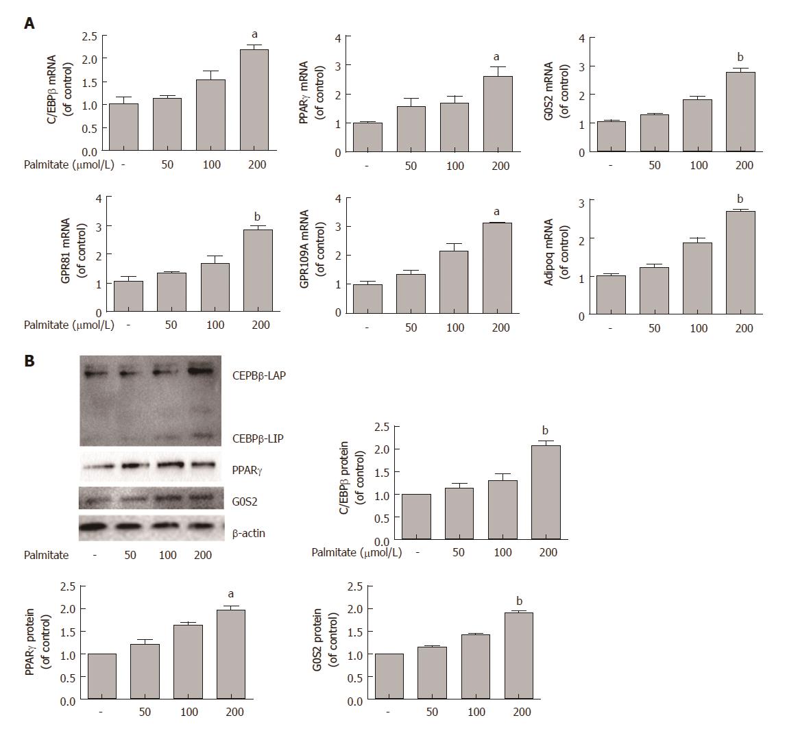
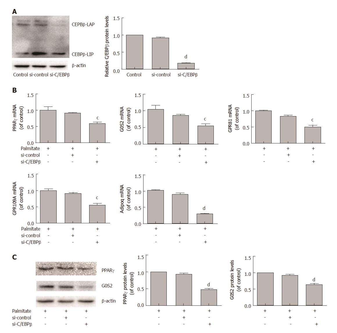
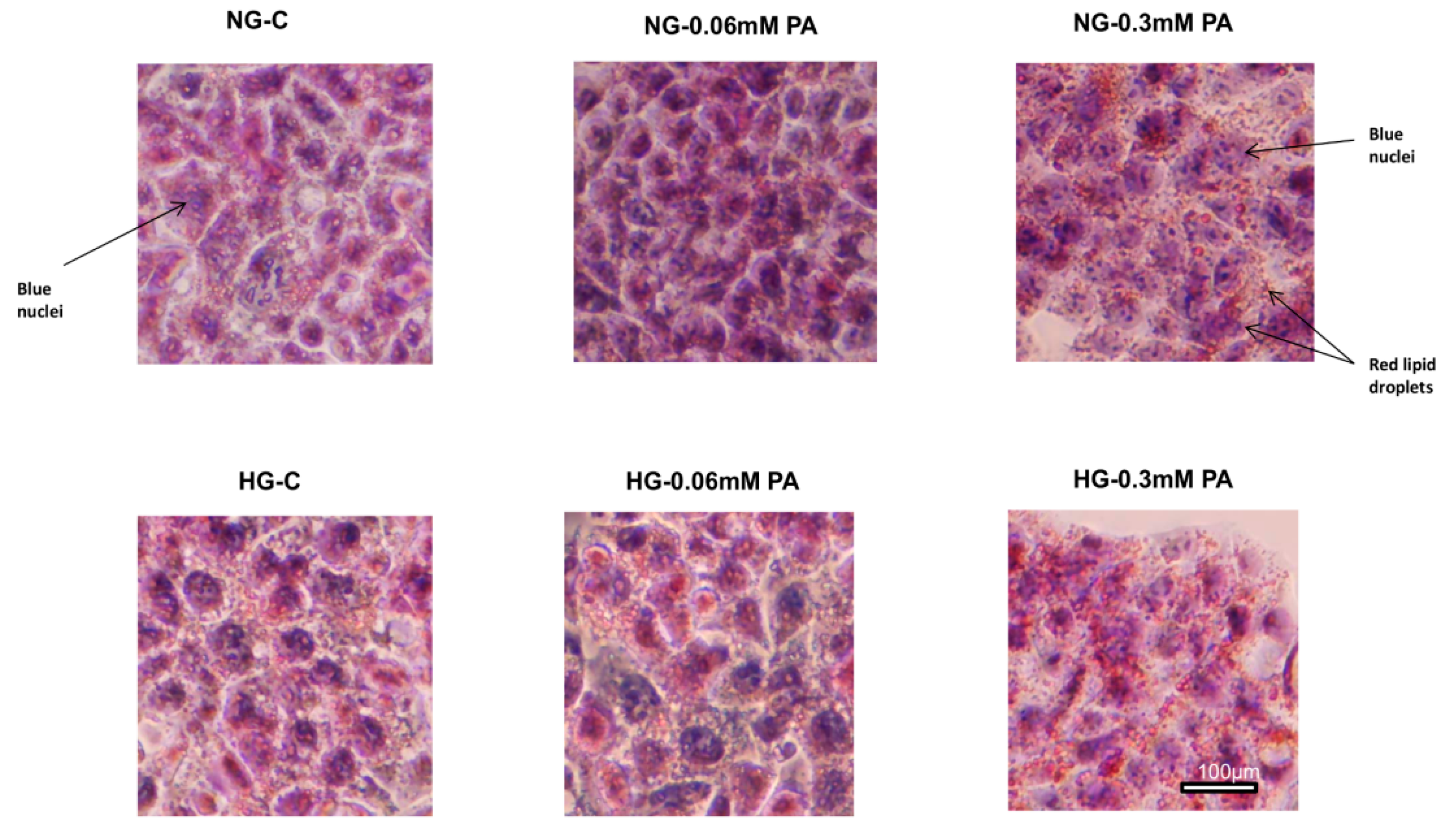
Oil red o staining hepg2 cells protocol- For Oil Red O staining, liver cryostat section (8 μm) or HepG2 cells were fixed and then were stained with Oil Red O (Solarbio, Beijing, China) according to the standard protocol For hepatic lipid quantification, approximately 100 mg of the liver tissue was homogenized in 02 mL of normal saline solution, and then hepatic lipids wereLipid accumulation was detected with Oil Red O staining and quantified by absorbance value of the extracted Oil Red O dye Lipolysis was evaluated by measuring the amount of glycerol released into the medium Results Palmitate caused a dosedependent increase in lipid accumulation and a dosedependent decrease in lipolysis in HepG2 cells

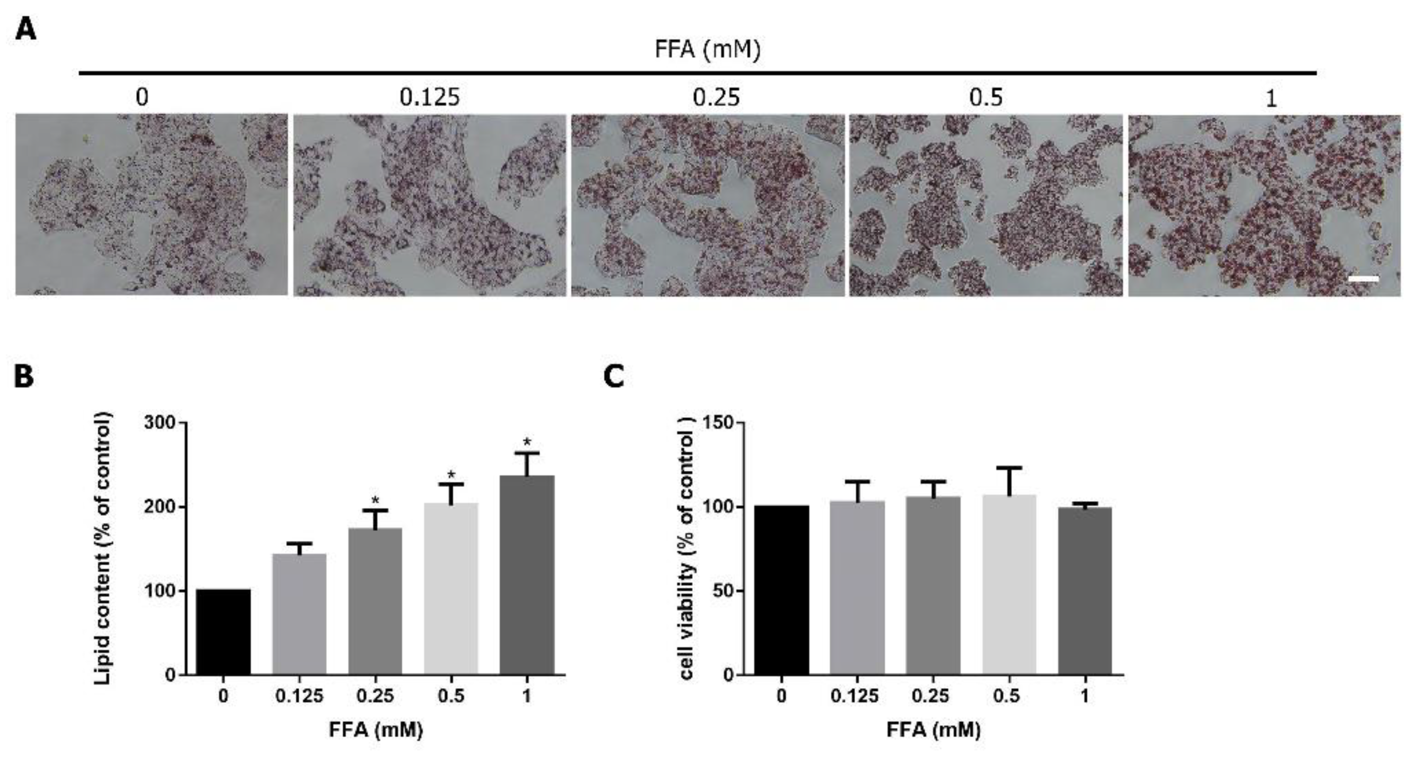
Lipid Accumulation In Hepg2 Cells By Oil Red O Staining After Treating Download Scientific Diagram
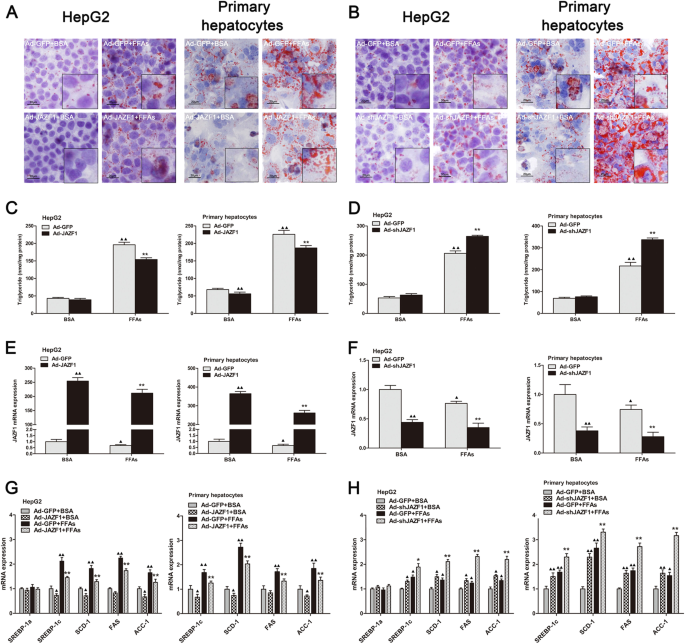
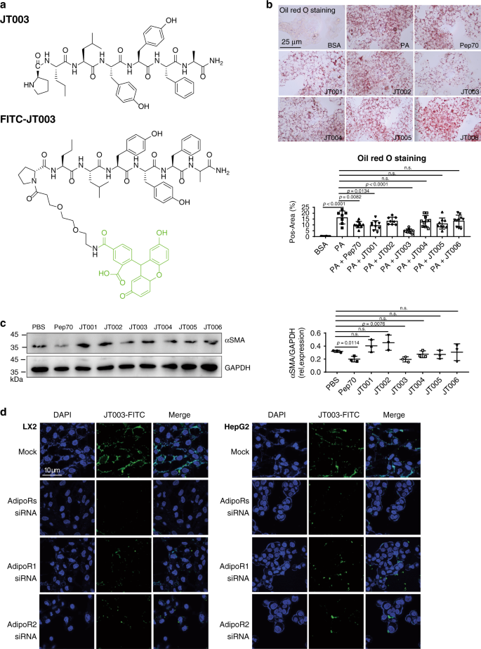
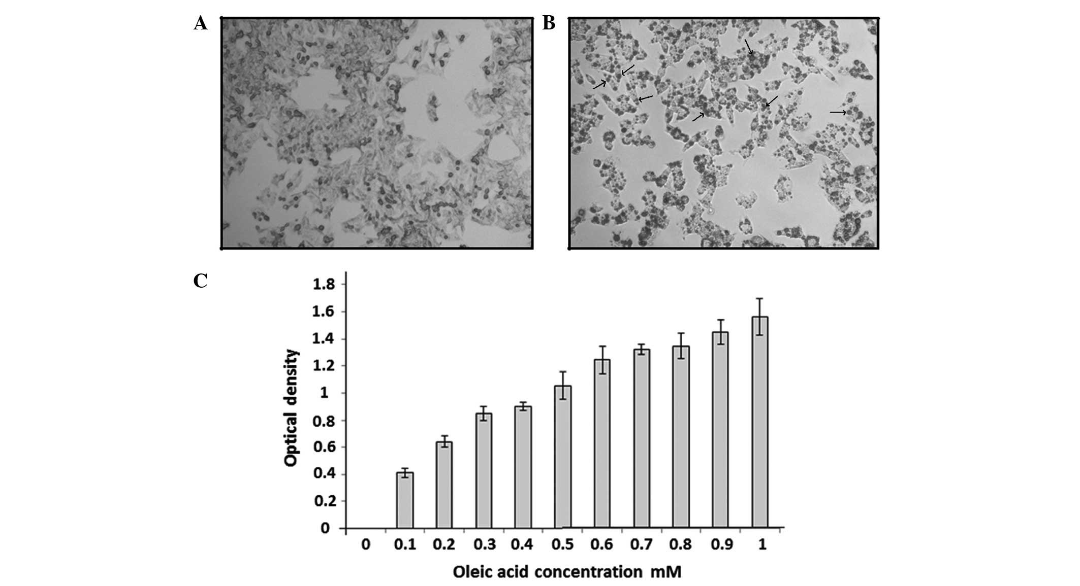
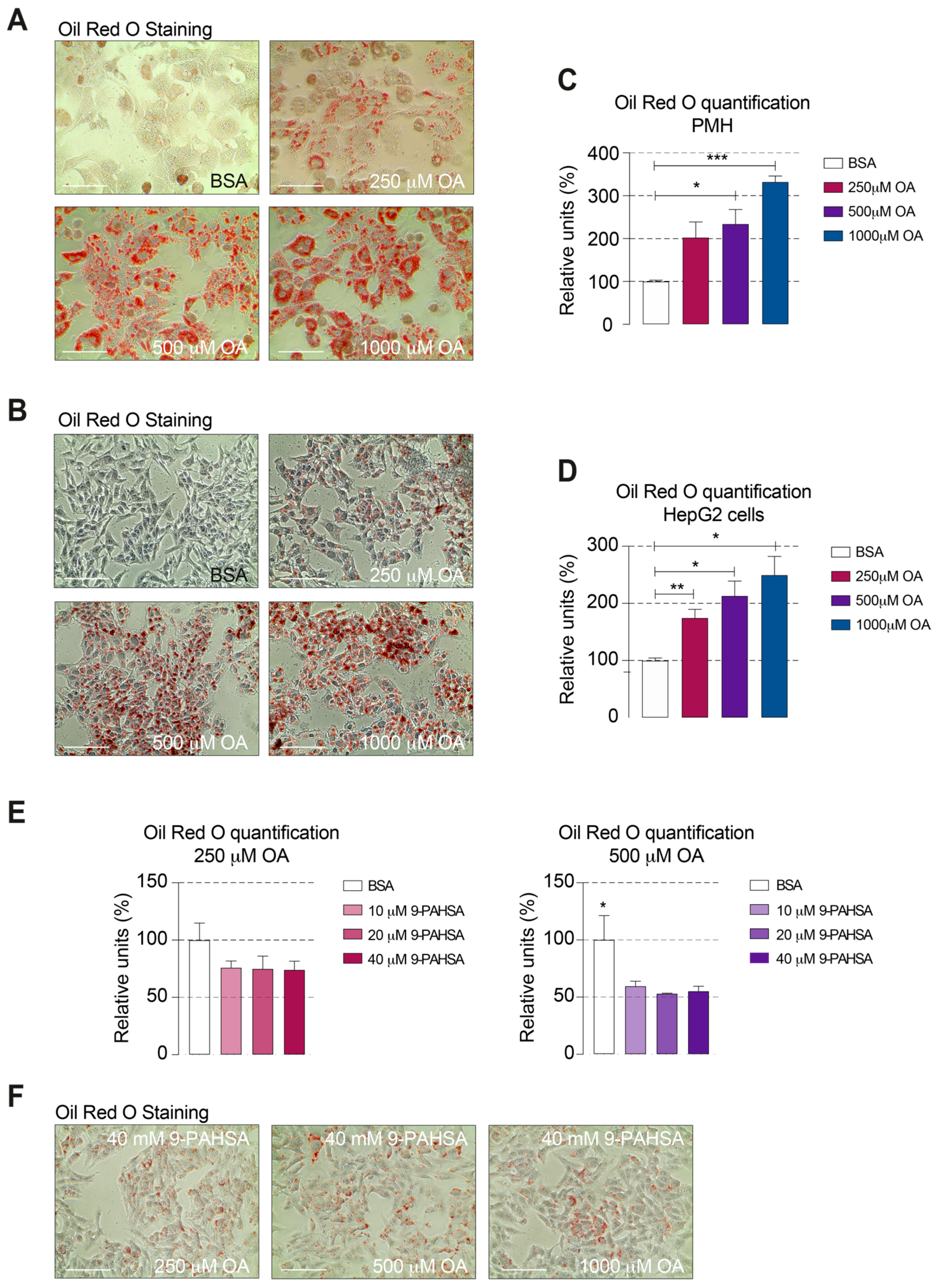
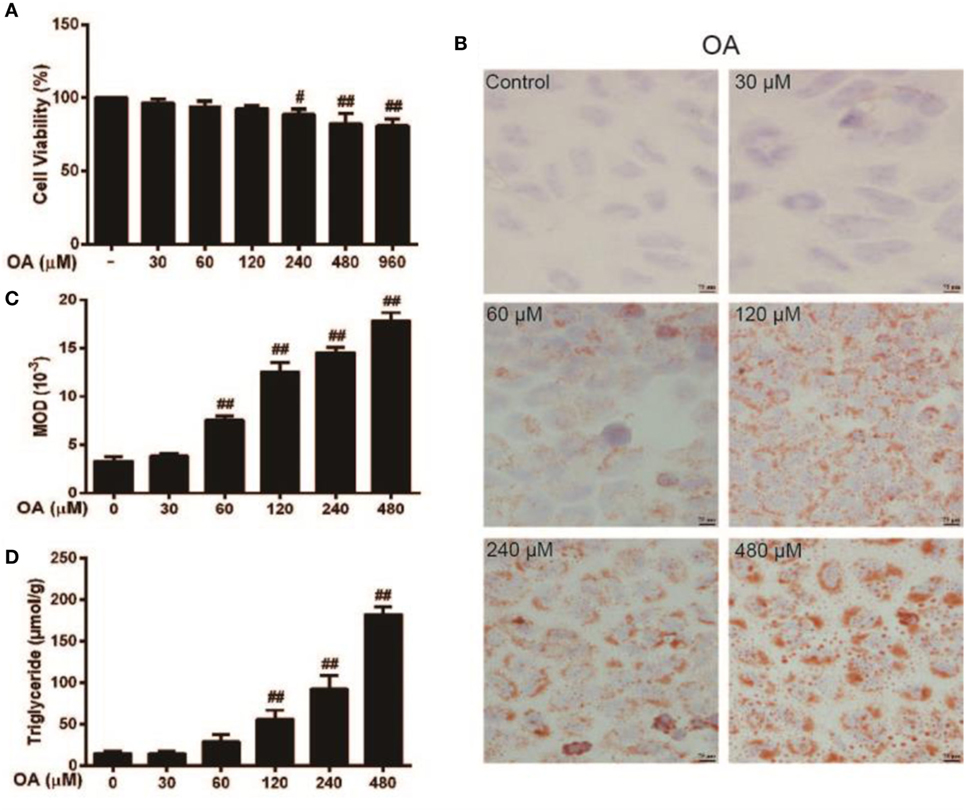
Materials and methods Cell culture treatment HepG2 cells, a human hepatoblastoma cell line, were culture in high glucose Dulbecco's modified Intracellular lipid content assessment HepG2 cells were plated in 25cm 2 flask at 70% confluence and coincubated with OilOil Red O staining (a) The effect of OA on hepatocyte steatosis in HepG2 cells The effects of RSV (b) and RSVPLGANPs (c) on TG accumulation in HepG2 induced by OA Source publication b Oil red O staining of HepG2 cells with adenovirus infection c Cellular triglyceride concentration analysis in HepG2 cells treated with adenovirus d h C57BL/6 J male mice (9 weeks old) were injected with AdGFP ( n = 8) or AdFoxO3 ( n = 10) through the tail vein at a concentration of 3 × 10 9 plaque forming units per mouse, fed a chow
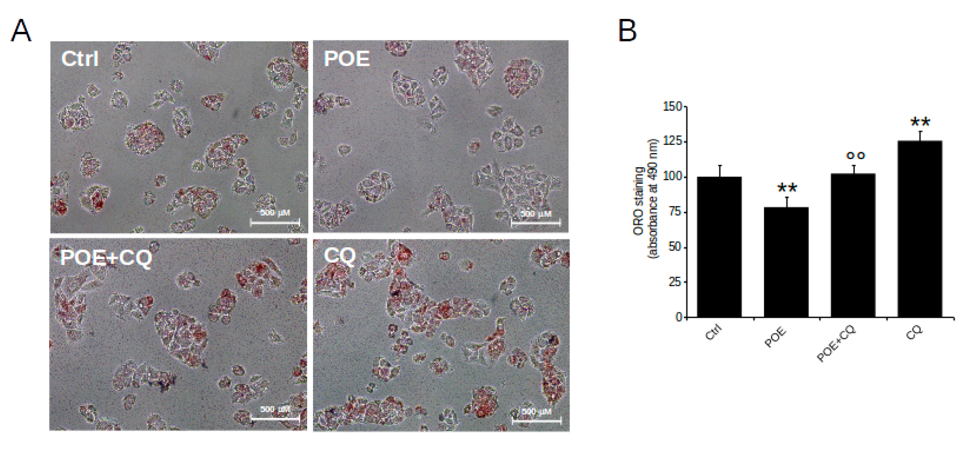
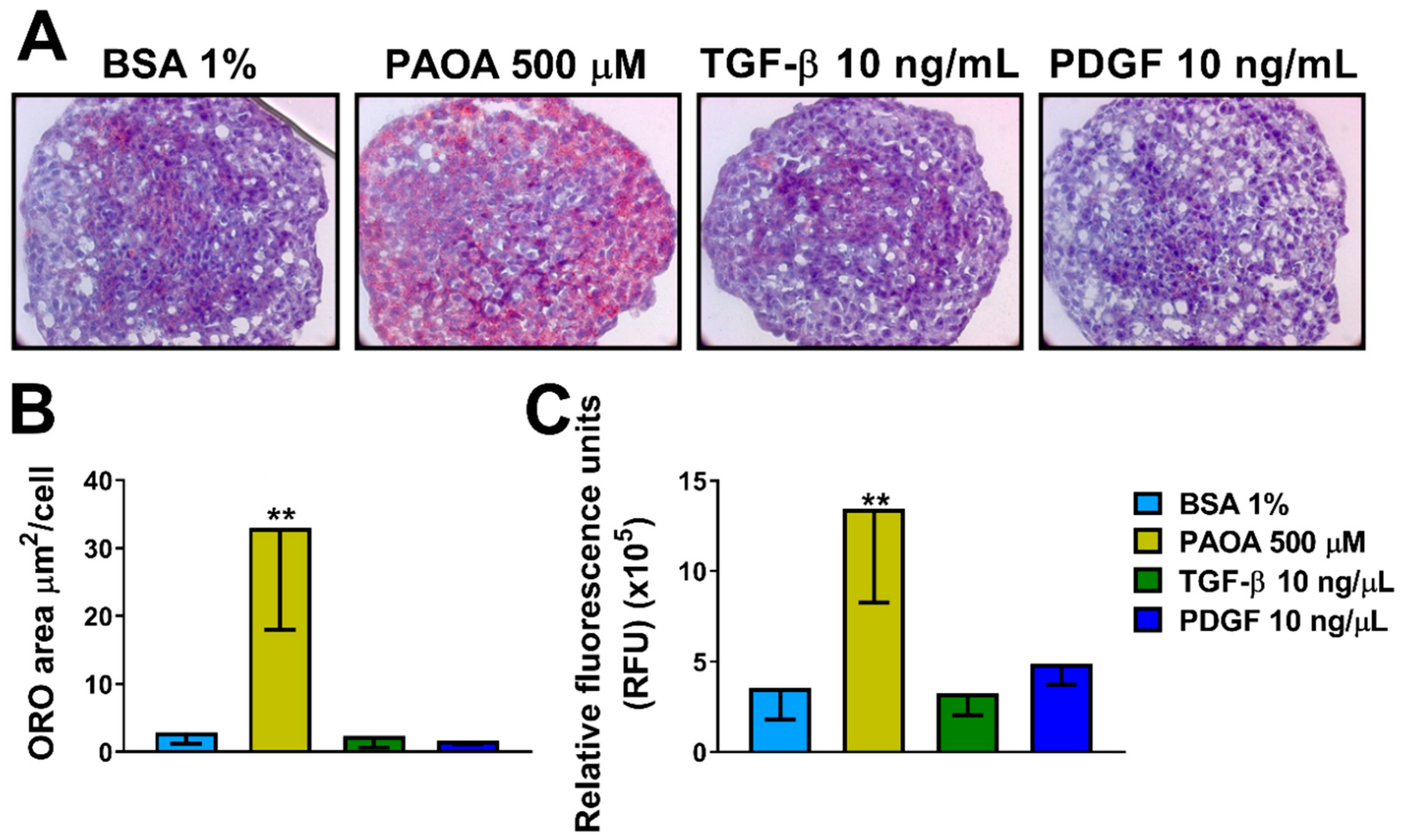
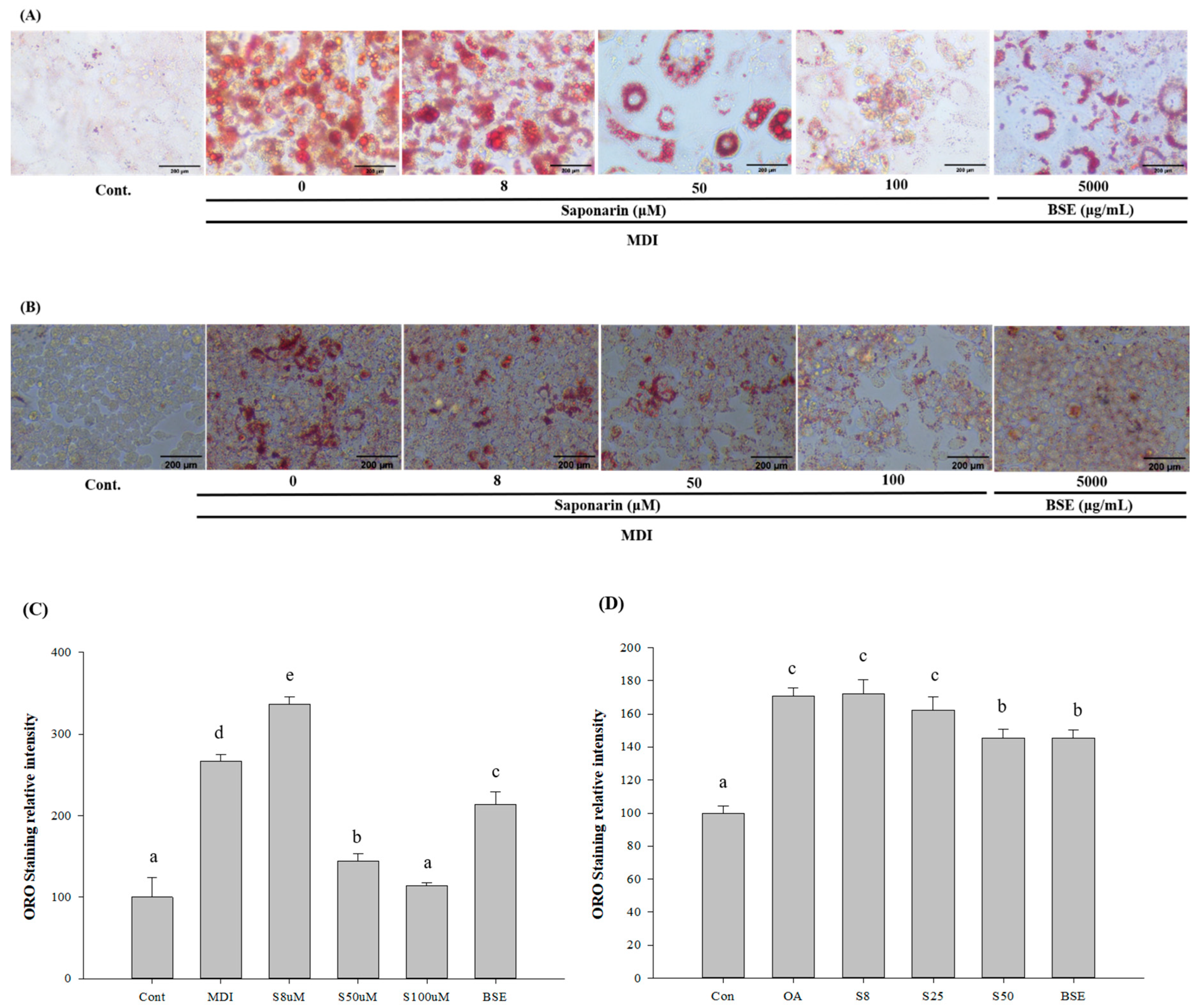
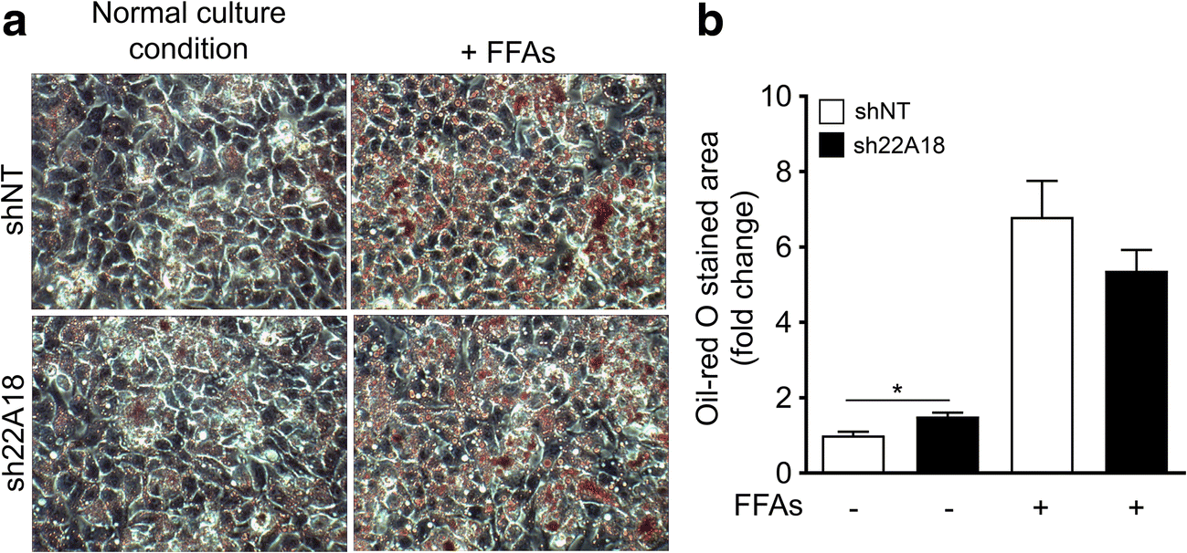
Main methods Oleic acid (OA) induced hepatic steatosis was established in L02 and HepG2 cells as in vitro model of NAFLD Cell apoptosis, lipid accumulation and oxide stress were evaluated by flow cytometry, oil red O staining, and cellular biochemical assays, respectivelyOilRedO Staining Subconfluent monolayers of HepG2 cells were exposed to palmitic acid or BSA as a control for 12 hours Cells were stained with OilRedO to examine the amount of fat accumulation in the cells Briefly,disheswerewashedwithcoldphosphatebuffered salineandfixedin10%neutralformalinAfter2changes ofpropyleneglycol,OilRed Oil Red O staining and the determination of triglyceride, malondialdehyde, and reactive oxygen species (ROS) contents proved the elevated lipid accumulation and oxidative stress by the mixture of BDE9 and HF in HepG2 cells, consistent in C57BL/6 mice Importantly, the action analysis showed the synergistic effect between BDE9 and HF
Oil red o staining hepg2 cells protocolのギャラリー
各画像をクリックすると、ダウンロードまたは拡大表示できます
 |  | |
 |  | |
「Oil red o staining hepg2 cells protocol」の画像ギャラリー、詳細は各画像をクリックしてください。
 |  |  |
 |  | |
 |  | |
「Oil red o staining hepg2 cells protocol」の画像ギャラリー、詳細は各画像をクリックしてください。
 |  |  |
 |  | |
 |  | |
「Oil red o staining hepg2 cells protocol」の画像ギャラリー、詳細は各画像をクリックしてください。
 |  |  |
 |  |  |
 |  |  |
「Oil red o staining hepg2 cells protocol」の画像ギャラリー、詳細は各画像をクリックしてください。
 |  | |
 | ||
 | ||
「Oil red o staining hepg2 cells protocol」の画像ギャラリー、詳細は各画像をクリックしてください。
 |  |  |
 |  | |
 |  |  |
「Oil red o staining hepg2 cells protocol」の画像ギャラリー、詳細は各画像をクリックしてください。
 |  |  |
 |  |  |
 |  |  |
「Oil red o staining hepg2 cells protocol」の画像ギャラリー、詳細は各画像をクリックしてください。
 |  | |
 |  |  |
 |  |  |
「Oil red o staining hepg2 cells protocol」の画像ギャラリー、詳細は各画像をクリックしてください。
 |  |  |
 |  | |
 |  | |
「Oil red o staining hepg2 cells protocol」の画像ギャラリー、詳細は各画像をクリックしてください。
 |  | |
 |  |  |
 |  | |
「Oil red o staining hepg2 cells protocol」の画像ギャラリー、詳細は各画像をクリックしてください。
 |  | |
 |  |  |
 |  | |
「Oil red o staining hepg2 cells protocol」の画像ギャラリー、詳細は各画像をクリックしてください。
 |  |  |
 |  |
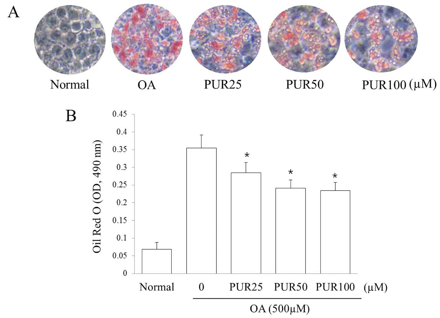
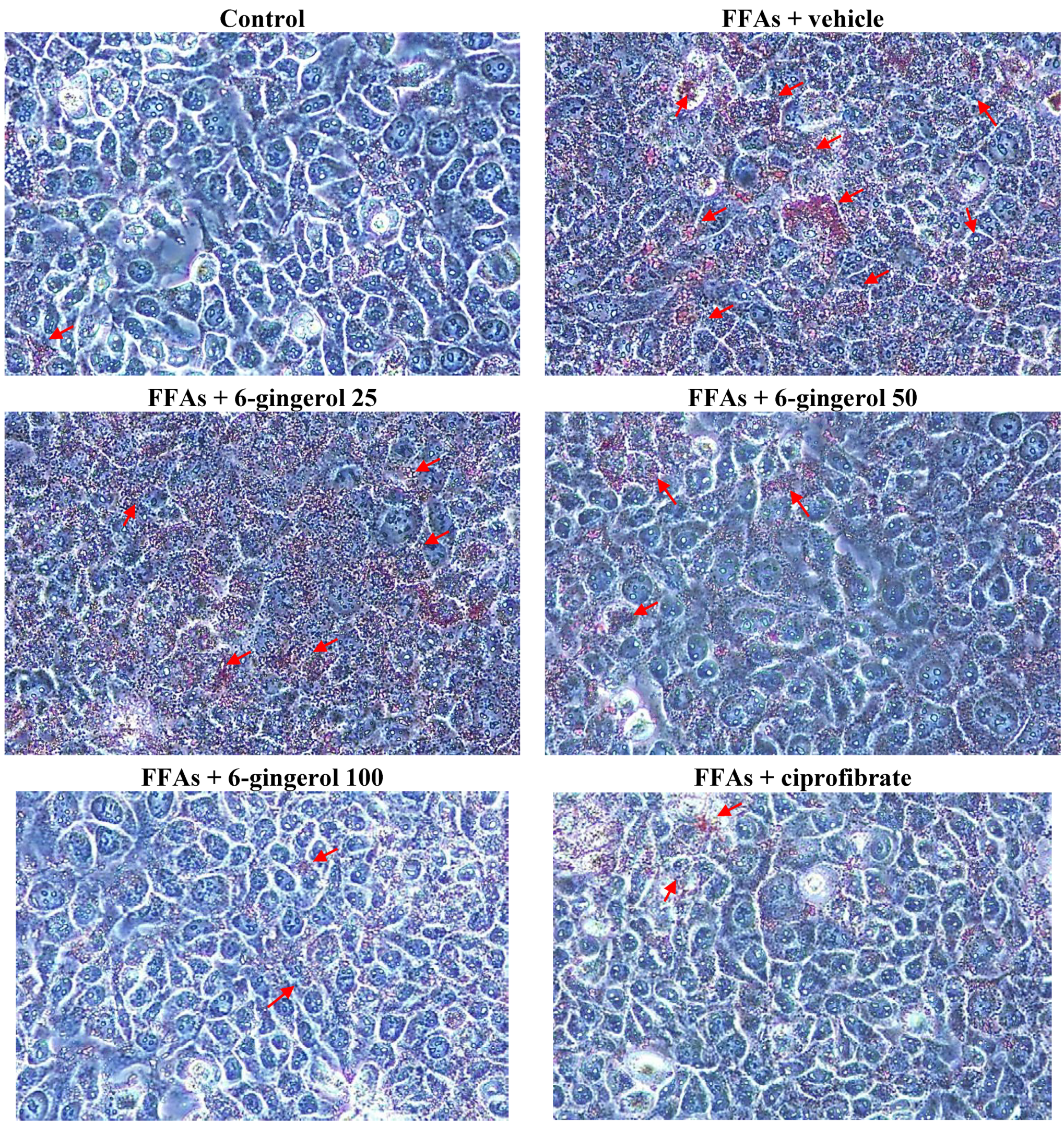
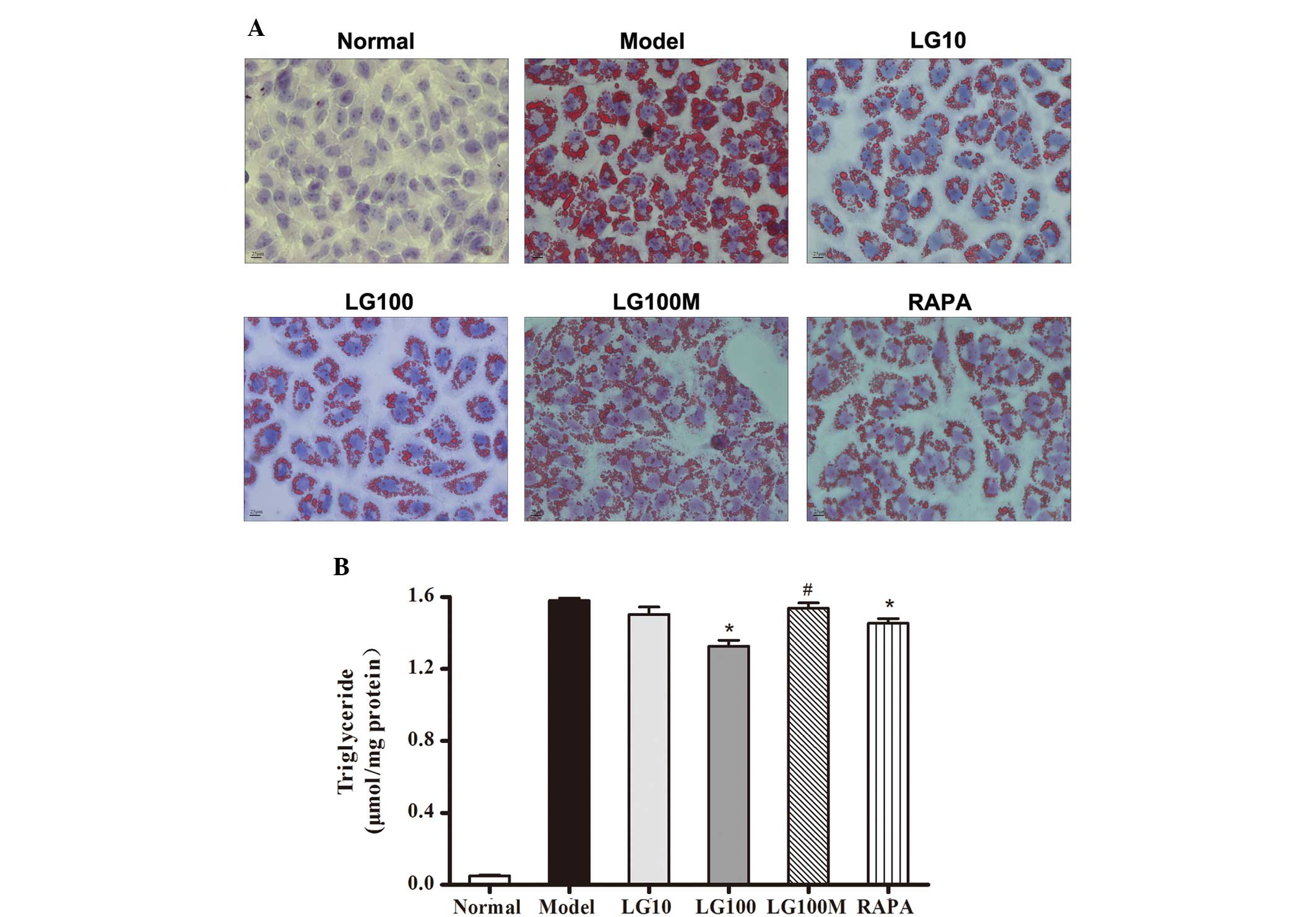
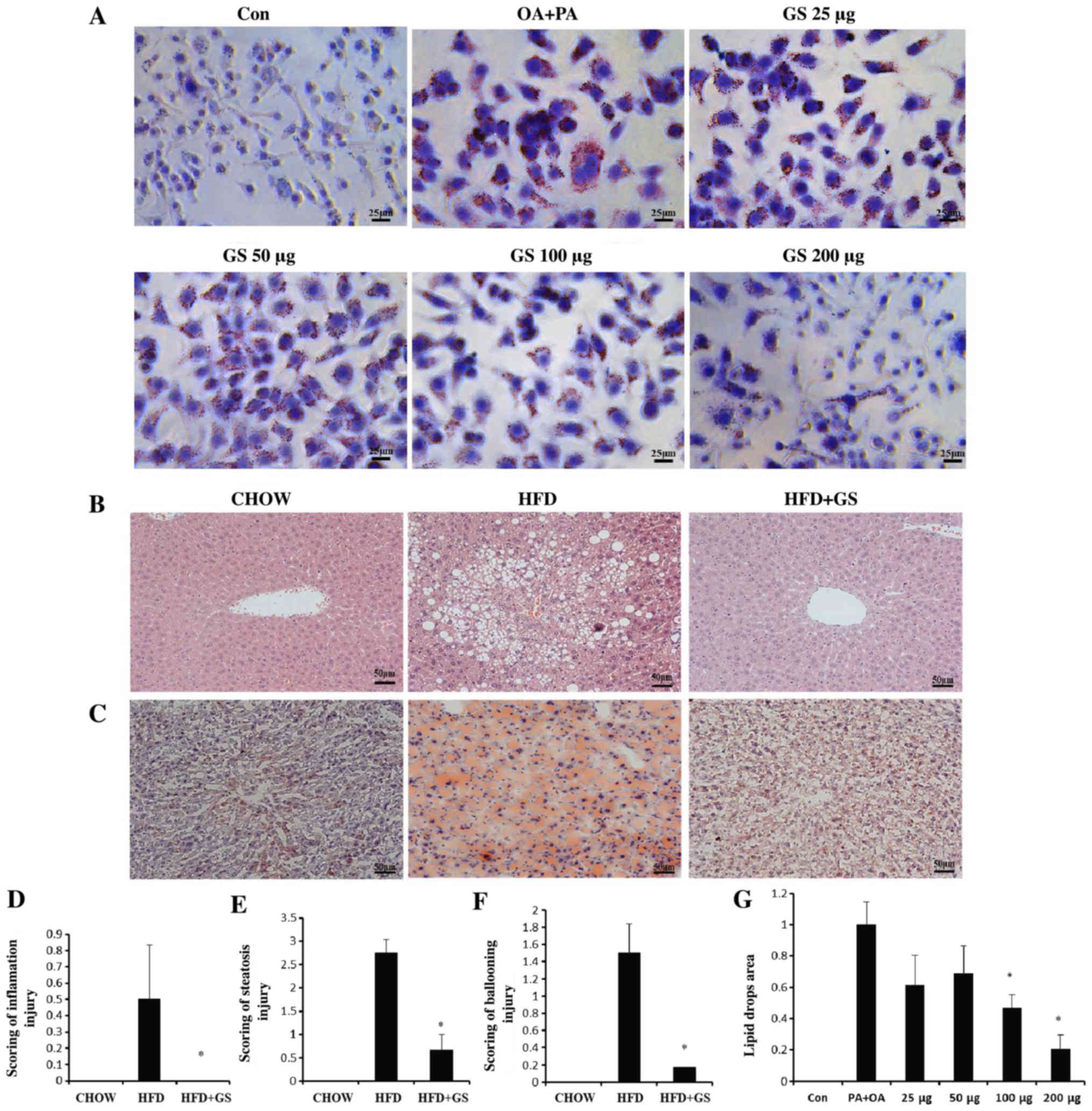
Treatment of HepG2 cells, a human hepatoblastoma cell line, with OA induces morphological similarities to steatotic hepatocytes 5, 6, but quantification of OAinduced steatosis has not been well established Oil red O (ORO) is a lysochrome (fatsoluble dye) diazo dye used for staining of neutral triglycerides and lipids on frozen sections Oil Red O staining and intracellular triglyceride levels showed marked accumulation of lipid droplets in both HepG2 cells and rat hepatocytes, and these were attenuated by GSO treatment In HFDfed mice, GSO improved HFDinduced dyslipidemia and
Incoming Term: hepg2 oil red o staining, oil red o staining hepg2 cells, oil red o staining hepg2 cells protocol,
コメント
コメントを投稿